10 FAQ About The Human Connectome Project, The Astonishing Sister Of NIH's Human Genome Project
At some point you may have wondered how exactly does your mysterious brain work. Maybe it was after you awoke from a strange and wonderful dream, or when you suddenly remembered a long forgotten word. While no one can truly answer how the brain works (yet), great progress has been made in recent years. One scientific endeavor in particular promises to deliver an abundance of information about this complex organ — the Human Connectome Project (HCP), a venture funded by the National Institutes of Health, is creating a map of the circuitry within the human brain.
The human what project, you say? Strangely, while the Human Genome Project snatched headlines for months, it seems few people have heard of its equally astonishing sister. This simple FAQ should help with that while also giving you something to talk about at Thanksgiving dinner.
What is a connectome?
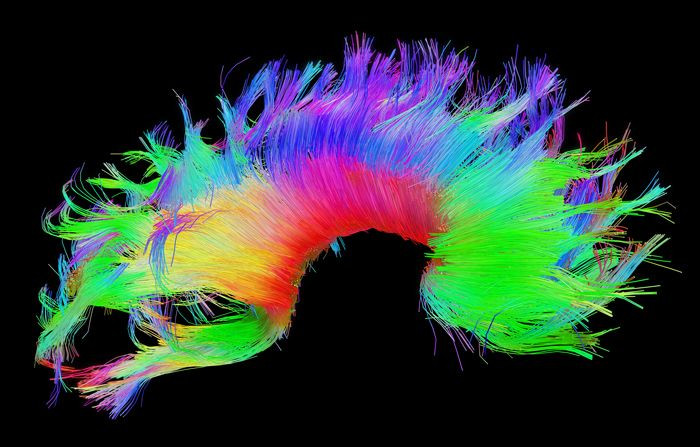
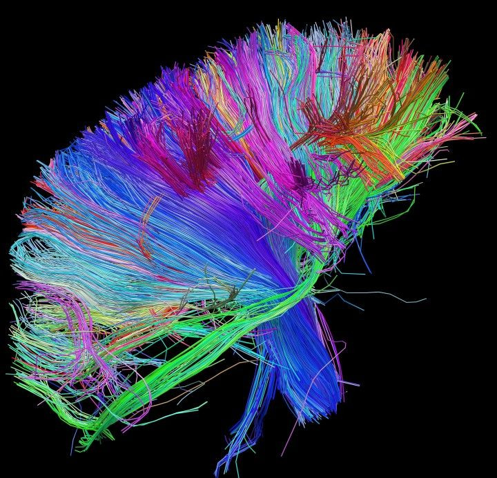
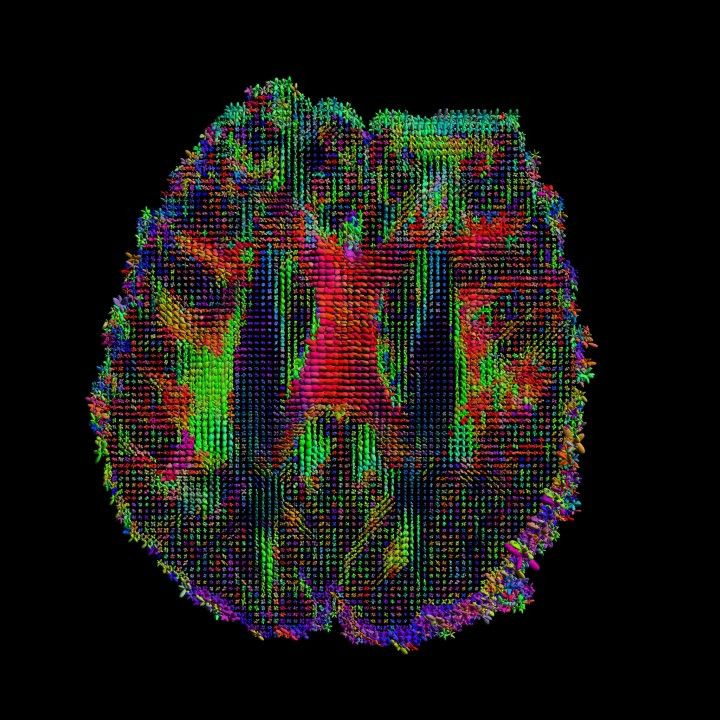
The suffix “-ome” refers to a totality, as The Scientist explains. So, just as a genome would refer to all of the DNA within a single cell, a connectome refers to all the cellular connections within the brain or, more simply, a connectome is a wiring diagram of the brain. In fact, each map reveals the architecture of our brain's white matter. More specifically, each diagram shows how bundles of fiber course through the white matter of the brain to form functional connections between gray matter regions. The fiber bundles look like “colored spaghetti,” Dr. Arthur Toga, a neuroscientist at USC’s Laboratory of Neuroimaging, Keck School of Medicine told Medical Daily.
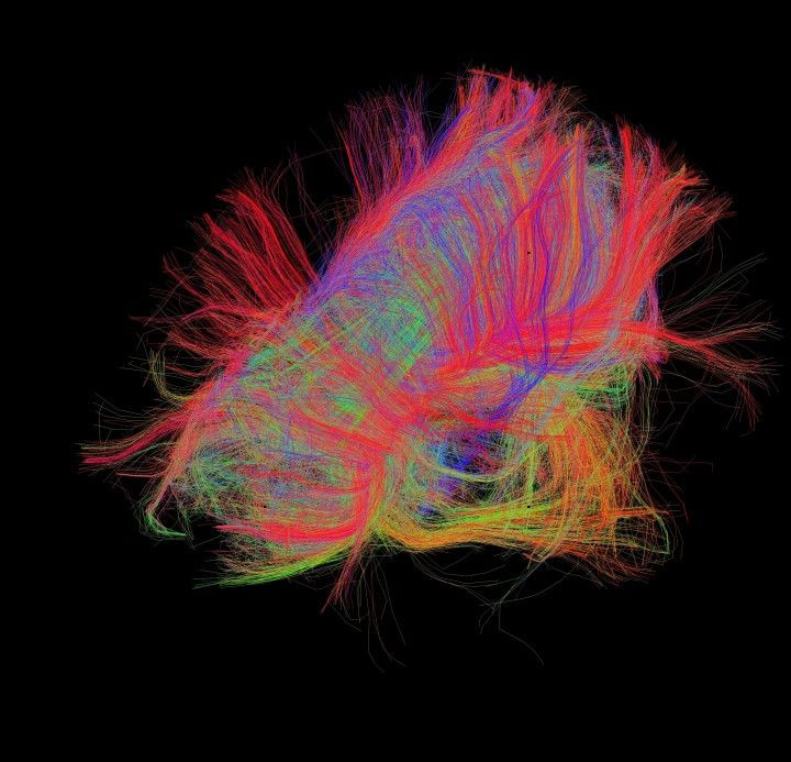
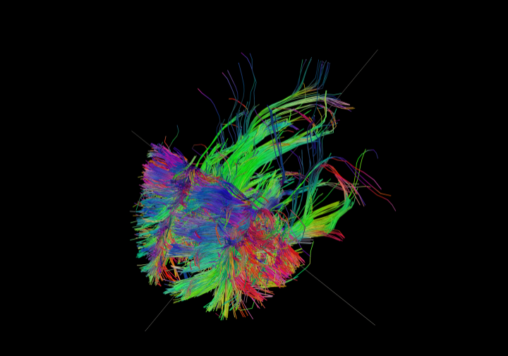
The colors in these images show the direction of the fibers: red = left-right, green = anterior-posterior, blue = through brain stem.
I’ve forgotten my brain basics. Can you remind me?
Medical Daily’s got your back. Our brains are mostly made of neurons, with scientists estimating their total number at 100 billion. These specialized cells of the nervous system are unique in that they have branching dendrites and axons, which serve as pathways allowing signals to travel from neuron to neuron. Information is sent by means of chemical and electrical pulses across conjunction sites referred to as synapses.
The brain’s outermost layer is made up of gray matter, which mostly contains neuron cell bodies. Tucked beneath the gray matter is white matter, which mostly consists of axons and glial cells, essentially support workers to the axons. White matter's main function is to facilitate communication between the neurons located in near and far regions of gray matter. Overall, the brain is an always pulsing, integrated network of short- and long-range connections between brain parts.
A much more extensive summary of brain basics can be found here.
Who is behind the Human Connectome Project?
The project comprises 36 investigators, including biologists, physicians, physicists, and computer scientists, at 11 institutions across the nation. The primary centers of research are USC’s Laboratory of Neuroimaging, Massachusetts General Hospital’s Martinos Center, Washington University’s Van Essen Lab, and the University of Minnesota’s Center for Magnetic Resonance Research.
The project was originally designed to generate rigorously collected data sets for distribution, “a rather unique collection,” Toga explained. “Ultimately, it stimulated unique advances in grouping data and analyzing data.”
What’s the project’s timeline and did I miss it?
The project was carried out in two phases. During Phase I, which spanned the years 2010 through 2012, research teams designed the project’s 16 major components. During Phase II, which ranged from 2012 through this past summer, the various scientists performed the actual work of gathering data. More importantly, however, during the most recent phase investigators made their datasets publicly available at regular intervals so that scientists around the world could begin to use them in their own projects.
The most likely reason you didn’t entirely miss the HCP is because research in this field is still advancing, with many scientists around the world continuing to use the project’s datasets.
Who helped the scientists? Who were the participants?
A total of 1,200 healthy adults, including a high proportion of twins and their non-twin siblings. This unusual emphasis on twins and their families was intended to help the researchers understand whether brain circuits are inherited. As part of their contribution, each participant had their genome mapped, allowing scientists to evaluate how much genes influence — or don’t influence — individual brain wiring. Each volunteer also completed a series of questionnaires and tests to measure behavior and demographic traits.
What kind of technology was used?
Only the coolest. To conduct the work, scientists used noninvasive neuroimaging equipment, including customized head coils and innovative MRI hardware. The high-resolution tools and technology are not only more sensitive but they also require less time to produce an image, compared to the past. Along with advanced computer science, the technologies reveal the brain as a whole, and at a level of detail not previously imagined possible in a living person.
What’s important to remember is this: Until very recently, it’s been difficult to study a living human brain. Much of our past knowledge of brain comes from research conducted on post-mortem samples, in vitro models, or animals.
What’s been discovered so far?
Many surprises. Scientists have been amazed to see that, instead of chaos, the connecting fibers are organized into an orderly 3D grid, where axons run up and down and left and right, minus any diagonals or tangles. Science magazine compares the brain’s 3D layout to New York City, with its streets running in two directions and buildings’ elevators running up and down. Strangely, in flat areas of the grid, the fibers overlap at precise 90 degree angles and weave together much like a fabric, the scientists say.
However, the bundles of fiber the project has mapped so far are really a 30,000-foot fly-by view, Toga said, continuing with the city metaphor. Though we can see the streets, highways, railroads, and other major transportation/communication lines, what we don’t see is how and where they connect to individual houses. “I believe it’s the connections to the house that really distinguish me from you and you from others,” Toga said.
Unique as each of our connectomes may be, similarities exist in the same way our physical architecture is roughly the same (we all have two legs, one nose, two eyes). Toga says different brain regions are different in some people early in life, while others change more over time due to greater plasticity.
Just as important, the twin studies demonstrated connectivity could sometimes be influenced by genetics, Toga said.
So this is it, or is there more?
Not by a long shot. Commentary on the project suggests both the technology and methodology, even if it is the most advanced in existence, had limitations that may have biased interpretation of the brain’s architecture. That said, the connectome is a moving target — the more we learn, the more there is to learn.
“The holy grail is to study connectomes as they change over time,” Toga said. Since some biomarkers of disease show themselves before deterioration begins, identifying the changes that occur before the onset of something like Alzheimer’s could lead to earlier therapy, and potentially stop or slow the disease’s progress, he explained.
Over time, technology will evolve, research will continue, and our understanding of the brain will become more nuanced, if never entirely complete.
Are there any recent connectome studies I could read?
Sure thing. This study, published in September and based on data from the HCP, revealed a strong relationship between positive behavior traits and the wiring of your brain. While some brains appear to be wired for a lifestyle that includes education and high levels of satisfaction, other brains appear to be wired for anger, rule-breaking, and substance use, according to the Oxford researchers.
Another study, based on HCP data and published earlier this month in Nature Neuroscience, demonstrates how our connectivity profiles are "intrinsic," acting "as a 'fingerprint' that can accurately identify subjects from a large group" regardless of how the brain is engaged during the scan. Though the researchers found connectivity patterns to be characteristic throughout the brain, the "frontoparietal network emerged as most distinctive." Connectivity profiles even predicted levels of fluid intelligence and cognitive behavior.
Read more from the ever-growing list of studies here.
In the end, what will be gained from the project?
In a previous email exchange about connectomics, Dr. Narayanan Kasthuri, assistant professor of anatomy and neurobiology at BU School of Medicine, said it best: “What parts of brain circuitry are stereotyped and which parts are variable across humans?”
“With a detailed connectome map of a normal human brain, I believe we will gain a better understanding of the roots of human neurological disorders, including schizophrenia, autism spectrum disorders, and other baffling conditions that may arise from abnormal ‘wiring’ during brain development,” wrote Dr. Francis Collins, director of the NIH, in a blog entry. He hopes this knowledge will ultimately help us learn how to detect, treat, and someday even prevent brain disorders.
