Autism Could Be Caused By Maternal Antibodies Targeting The Fetus

The myriad causes of autism are the subject of much research and scrutiny. Recent research focuses on events that occur during development before birth in order to explain how autism and autism spectrum disorders (ASD) occur. It has been established that autism and ASD are caused by alterations in brain development.
A new study at the University of California, Davis's MIND Institute has identified a maternal autoantibody that acts on the fetus to begin this alteration in brain development that will ultimately lead to autism or ASD.
Autism and ASD are characterized as having difficulty with social and emotional exchanges and deficits in nonverbal communication used for social interaction. Similarly, people with ASD have difficulty developing and maintaining relationships with others.
Antibodies, or immunoglobulins (Ig), are a crucial part of the body's immune system. They act to identify and help the body get rid of foreign, disease-causing agents like bacteria and viruses. But sometimes, the body can make autoantibodies, which target the body's own cells for destruction. This can cause autoimmune diseases like psoriasis or type I diabetes, where the immune system destroys cells necessary to the body.
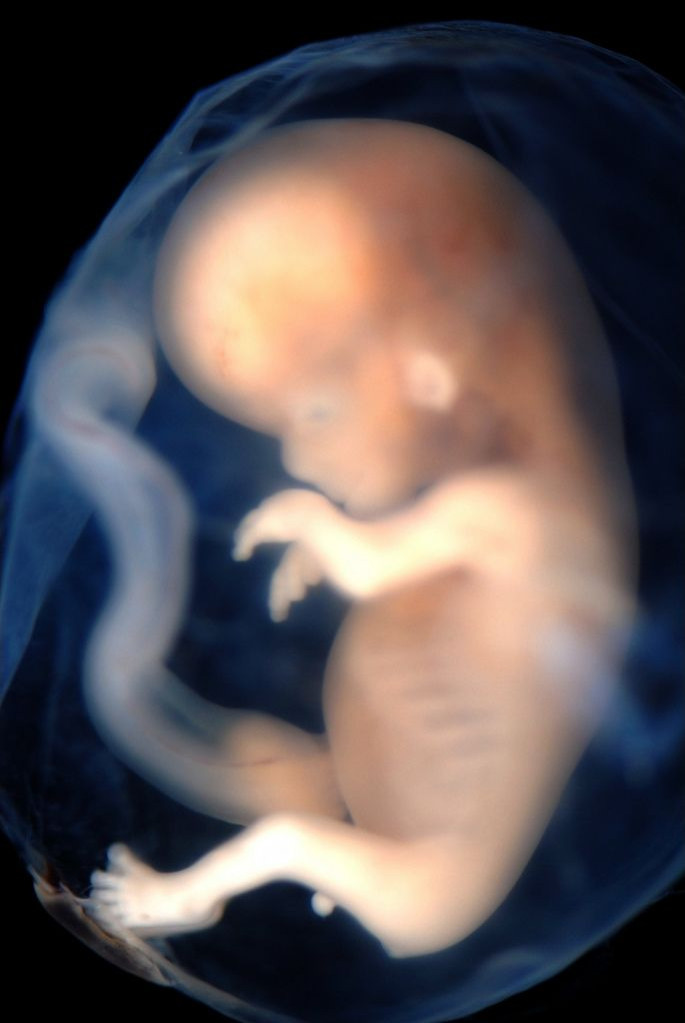
Immune disorders can be particularly dangerous during pregnancy, as the placenta is the only barrier between the fetus and its mother's antibodies. Often, the mother's antibodies will cross the placenta and protect the fetus from infections. However, if autoantibodies cross the placenta, they do not protect, but rather attack the fetus. The particular autoantibody, called IgG, found to cause ASD is present in 12 percent of women who have children who develop ASD. IgG specifically attacks the fetus' brain when it crosses the placenta.
The IgG's preference for brain tissue is of interest, as ASD is known to be caused by brain developmental deficits. Researchers collected blood serum from mothers of children with and without ASD. The mothers of children with ASD had the IgG, which was purified to be used. Rhesus monkeys were used in this study as they have similar brain organization and cognitive and social functions as humans. Three groups were used, each composed of eight monkeys: one group was given the IgG serum from mothers of children with ASD, another was given the serum from mothers with children without ASD, and a third group was left untreated.
After birth, baby monkeys were given longitudinal magnetic resonance imaging scan (MRI) at six months of age to check for physical developmental differences in their brains at one week, one month, three months, six months, one year, and two years of age. The animals were also observed for changes in behavior and social development, as ASD is often characterized as an inability to develop social norms.
Differences seen among the monkeys who had received the ASD-related IgG (IgG-ASD), compared to the monkeys without IgG-ASD and the control group, were telling. Brain growth was significantly faster in the IgG-ASD monkeys, and they had bigger brains than the control animals by the age of two. The bigger brains were also organized differently from normal brains; an increase in white matter over grey matter is often indicative of autism in humans.
However, behavioral data revealed more. When faced with different social situations, the IgG-ASD monkeys behaved in a way that indicated they had or would soon develop autism. When faced with another monkey in a cage, all the monkeys tested chose to spend time with the other monkey. However, the IgG-ASD monkeys only spent an average of three seconds in the cage with the other monkey before returning to an empty cage. The IgG-ASD monkeys were also found to refuse companionship; the monkeys would approach others, but would not do so more than once or sustain interactions with any one monkey.
What was most interesting was that the IgG-ASD animals tended to approach others more frequently than expected. This is also deviant behavior for animals with social disorders. When dealing with a disorder like autism, animals tend to keep to themselves or have strained interactions with others. Researchers found that as the IgG-ASD monkeys got older, they approached other monkeys more than usual, but did not interact by playing with them.
"The combination of brain and behavioral changes observed in the nonhuman primate offspring exposed to these autism-specific antibodies suggests that this is a very promising avenue of research," said Melissa D. Bauman, Ph.D., lead author of the study, "highlighting the collaborative efforts that characterize research at the UC Davis MIND Institute."
Behavioral and MRI results from this study indicate that the human IgG-ASD antibody from mothers of children with ASD can create abnormal development in others if the antibody is given during fetal development. The antibody disrupts brain development, and is a potential cause of some forms of autism. While the monkeys who received IgG-ASD developed a form of autism, more work needs to be done to establish an effect of the maternal IgG in humans at different parts of development.
"Much research remains ahead of us to identify the mechanisms by which the antibodies affect brain development and behavior. But, this program of research is very exciting, because it opens pathways to potentially predicting and preventing some portion of future autism cases," said David Amaral, research director at the MIND Institute and senior author of the paper.
Source: Bauman MD, Iosif A, Ashwood P, et tal. Maternal antibodies from mothers of children with autism alter brain growth and social behavior development in the rhesus monkey. Translational Psychiatry. 2013.
Published by Medicaldaily.com


