Brain Images Indicate Biomarkers For Autism Spectrum Disorder, May Revolutionize Treatment

Autism is now one of the fastest-growing developmental disorders in America, affecting one in 68 children, for whom no known cure or objective diagnosis exists. A new study published in JAMA Psychiatry may amass hope for those affected by autism spectrum disorders, thanks to a new method for mapping the brain using functional magnetic resonance imaging (fMRI). The ability to identify a marker in brain scans that can be used to diagnose and measure the progression of autism would drastically change interventions among children.
"This is significant because biomarkers give us a 'why' for understanding autism in boys that we haven't had before," said the study’s co-author Dr. Kevin Pelphrey, director of the Autism and Neurodevelopmental Disorders Institute at George Washington University, in a statement. "We can now use functional biomarkers to identify what treatments will be effective for individual cases and measure progress."
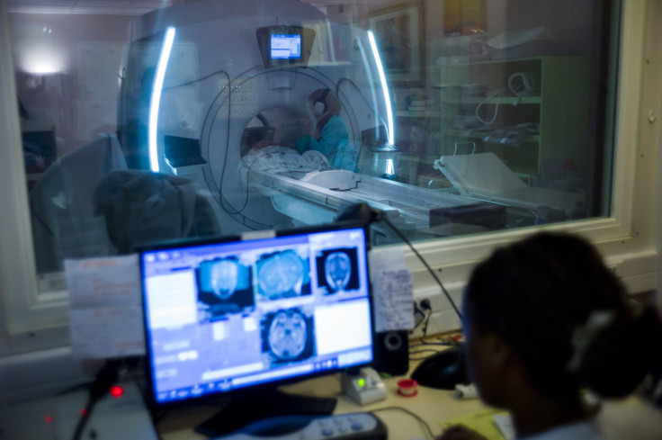
For the study, researchers collected 164 images from brain scans of 114 individuals using fMRI, which reveals changes in the brain as they occur. The scans revealed that boys on the spectrum had changes in a region in their brain responsible for social perception. Boys are five times more likely to be diagnosed with autism compared to girls, yet it wasn’t until now that researchers identified what makes their brain regions different than those of girls.
Having a skewed social perception, even a subtle one, may lie at the root of the gender differences. Difficulty communicating in social interactions and repetitive behaviors are some of the key characteristics of autism, which makes identifying a marker in the brain integral to unlocking a treatment.
"Brain function markers may provide the specific and objective measures required to bridge this gap," said the study’s lead author Malin Björnsdotter, assistant professor at the University of Gothenburg, in a statement. “To really help patients, we need to develop inexpensive, easy-to-use techniques that can be applied in any group, including infants and individuals with severe behavioral problems. This study is a first step toward that goal."
Not only will the brain scans help with diagnosis, they will also serve as a tool for measuring progress during treatment. Doctors will get a clearer and more definite diagnosis while also having the opportunity to develop a more tailored and effective treatment plan for the child, whether it's a behavioral or drug intervention, or a combination of the two. Doctors could use fMRI scans to see in real time if a particular treatment helps to improve symptoms, or even treat the disorder itself.
"This kind of imaging can help us answer the question, 'On day one of treatment, will this child benefit from a 16-week behavioral intervention?'" Pelphrey said. "Answering that question will help parents save time and money on diagnosis and treatments."
For their next step, Pelphrey and his team plan to study a larger population of patients with autism, along with those who have other neurological disorders. This way, researchers will determine if they can distinguish autism spectrum disorders from other disorders. If they can, then they'll be able to measure the symptoms and track treatment.
Source: Björnsdotter M, Pelphrey K, Wang N, and Kaiser MD. Evaluation of Quantified Social Perception Circuit Activity as a Neurobiological Marker of Autism Spectrum Disorder. JAMA Psychiatry. 2016.


