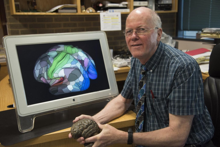
New Brain Map Marks Nearly 100 Unmapped Regions, Helps Scientists Better Understand Cerebral Cortex

Humans have designed maps for nearly every corner of the world, and we can now track different locations via Google. But in the modern age, our external explorations have turned inward, and scientists are now attempting to map out the most complicated human organ: the brain.
In a new study out of Washington University School of Medicine in St. Louis, a research team has developed an extensively detailed map of the brain’s cerebral cortex, the outer layer and the area involved in sensory perception, attention, language, tool use, and abstract thinking. The study, published in Nature, aims to not only better understand the brain’s hardware, but also to help in future research for brain disorders like dementia, autism, and schizophrenia.
“The brain is not like a computer that can support any operating system and run any software,” said David Van Essen, professor of neuroscience and an author of the study, in a press release. “Instead, the software — [or] how the brain works — is intimately correlated with the brain’s structure — hardware, so to speak. If you want to find out what the brain can do, you have to understand how it is organized and wired.”

For many years, scientists referred to a brain map that had been developed by Korbinian Brodmann, a German neuroanatomist, in the early 1900s. But after a while, researchers began to feel the need for an upgraded version of the map; one that could provide more information about its complexity. “My early work on language connectivity involved taking that 100-year-old map and trying to guess where Brodmann’s areas were in relation to the pathways underneath them,” said Matthew Glasser, lead author of the new study, in the press release. “It quickly became obvious to me that we needed a better way to map the areas in the living brains that we were studying.”
To create the map, the researchers used data from the Human Connectome Project, which has used a robust MRI machine to map out the brains of 1,200 young adults. They focused specifically on 210 healthy young adults, both male and female, and pooled a lot of data on their brains — including information about the thickness of the cortex, the insulation around neuronal cables, and MRI scans of the brain at rest and at work. The map delineated the brain regions in more detailed and precise ways, first dividing the left and right cerebral hemisphere into 180 areas based on physical, functional, and connective differences. Then they laid out the cortex, which consists of neural tissue and gray matter and is the outer layer of the brain. All of this involved an algorithm that can identify the regions of people’s unique brains.
"We ended up with 180 areas in each hemisphere, but we don't expect that to be the final number," said Glasser in the press release. "In some cases, we identified a patch of cortex that probably could be subdivided, but we couldn't confidently draw borders with our current data and techniques. In the future, researchers with better methods will subdivide that area. We focused on borders we are confident will stand the test of time."
Such detailed brain maps could help doctors personalize treatments for neurological disorders or mental illnesses, like Alzheimer’s. The way the researchers see it, it’s now a time of exploration inwards, and they believe their maps will help scientists around the globe to learn more about the brain. “We think it will serve the scientific community best if they can dive down and get these maps onto their computer screens and explore as they see fit,” said Van Essen in the press release.
Source: Glasser M, Coalson T, Robinson E, Hacker C, Harwell J, Yacoub E. A Multi-Modal Parcellation of Human Cerebral Cortex. Nature, 2016.
Published by Medicaldaily.com


