Food Imagery Helps Doctors Learn: Describing Diseases Using Food Enhances Doctors’ Diagnostic Abilities

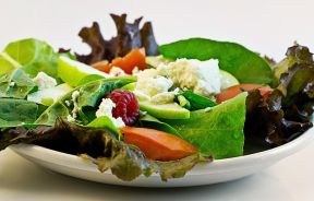
Describing medical conditions using food may sound strange at first, but “beer bellies” and “port wine stains” have become staples in the medical community and terms so commonly used, most don’t give it a second thought. Using food as visual and diagnostic tools provides a more thorough understanding of the condition or disease, argues a UK pathologist in Medical Humanities.
"It is a wonder that, in the midst of the smells and sights of human affliction, a physician has the stomach to think of food at all," said Dr. Ritu Lakhtakia, Departmentof Pathology, College of Medicine & Health Sciences, Sultan Qaboos University.
The human preoccupation with food has made it easy to turn tumors into mushrooms and cauliflower florets with word associations. There are a wealth of allusions to raw and cooked food and drinking in the medical field, and Lakhtakia argues it could also deter some patients and doctors from the food itself. However, it has become such a useful aid for doctors to remember complex conditions and unpronounceable medical terms.
After generations of food aromas, shapes, colors, and textures laced their way through medical writing, the food imageries are beginning to decrease from use, according to the paper. The use of food imagery in the health field is considered a powerful tool, which should secure its place in both medical teaching and records.
"Whatever the genesis, these time honored allusions have been, and will continue to be, a lively learning inducement for generations of budding physicians," Lakhtakia said.
The Most Well-Known Medical Food Imageries:
1. Beer Belly
A visually bloated stomach of abdominal fat caused by excessive consumption of beer and other high-carb or high-fat foods. It also generally describes obese conditions typical of males.
2. Port Wine Stain
A reddish-purplish, flat birthmark caused by swollen blood vessels that may darken as children get older.
3. Spaghetti and Meatball Appearance
Tinea versicolor is a fungal infection of the skin of yeast and hyphae, which can be seen on a vaginal smear on a microscopic slide.
4. Apple or Pear Shape
Describes the fat distribution of two different body types, the apple being unhealthily rounded in the middle and the pear-shape being more bottom heavy.
5. Milk Patch
Rheumatic pericarditis develops whitish plaques caused by the inflammation of the heart’s membranes.
6. Croissant Appearance
Schwannoma is a noncancerous tumor that develops on the peripheral nerves, and the nucleus of the spindle cell tumor resembles a croissant.
7. Blueberry Muffin Rash
Congenital rubella occurs when a mother has rubella virus in the first three months of pregnancy, during which the baby develops blue-grey nodules beneath the skin.
8. Anchovy Sauce
Amoebic liver abscess causes the excretion of dark brown pus.
9. Cottage Cheese Appearance
Mucosal candid infections develop curd-like resemblances because of its white granular appearance.
10. Strawberry Cervix
Trichomonas infection causes an inflamed, red appearance of the cervix.
11. Cherry Red Spots
Cherry Angioma or Campbell de Morgan spots are skin angioma characterized by their red spots and is often found on older people or those with lipid-storage disorders.
12. Chocolate Cyst
Endometriotic cyst on the ovary contains dark brown fluid from repeated cycles of uterine wall shedding and hemorrhaging.
Published by Medicaldaily.com


