New Microdevice Enables Culture of Rare Circulating Tumor Cells from Blood

A research collaboration between the Wyss Institute for Biologically Inspired Engineering at Harvard University and Children’s Hospital Boston has created a microfluidic device that can harvest rare circulating tumor cells (CTCs) from blood to enable their expansion in culture for analysis. These cells, which have detached from a primary cancer site and often create a secondary -- or metastasized -- tumor, hold an extraordinary amount of information regarding patient-specific drug sensitivity, cancer progression, and patient response to therapy.
Such information could help clinicians treat patients, but it has not been easily accessed due to the difficulty of isolating CTCs and expanding them in culture for subsequent analysis. In alleviating this problem, the new technology has the potential to become a valuable tool for cancer diagnosis and personalized treatment. The research findings appear online in the journal Lab on a Chip.
Wyss Founding Director, Donald Ingber, M.D., Ph.D., and Wyss Postdoctoral Fellow Joo Kang, Ph.D., led the research team. Ingber is the Judah Folkman Professor of Vascular Biology at Harvard Medical School (HMS) and the Vascular Biology Program at Children's Hospital Boston, and Professor of Bioengineering at Harvard's School of Engineering and Applied Sciences. Kang is a Research Fellow at Children’s Hospital. Also on the team were Wyss Postdoctoral Fellow Mathumai Kanapathipillai; Children’s Hospital Research Fellow Silva Krause and Research Associate Heather Tobin; and Akiko Mammoto, an Instructor in Surgery at HMS and Children’s Hospital.
This novel approach for capturing and culturing CTCs combines micromagnetics and microfluidics within a cell-separation device, about the size of a credit card, in which microfluidic channels have been molded into a hard clear polymer. As blood flows through these channels, magnetic beads that have been coated to selectively stick to the CTCs are used to separate them from the other cells in the blood. The dimensions of the channels have been designed to protect CTCs from mechanical stresses that might alter their structure or biochemistry, as well as to maximize the number of CTCs that can be captured.
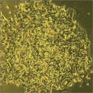
In the lab, the new approach demonstrated extremely high efficiency by capturing more than 90 percent of CTCs from the blood of mice with breast cancer. Of particular significance was the fact that the captured CTCs were able to be grown and expanded in culture. These intact living tumor cells could be used for additional testing and molecular analysis, for example, in screening drugs to meet the personal needs of individual patients in the future. Further testing found that the device is sensitive enough to detect the sudden increases in the number of CTCs that signal a cancer’s metastatic transition and could therefore alert clinicians to possible disease progression.
The Wyss Institute/Children’s Hospital team carried out their studies with one common type of breast cancer. But the same device could be used to address a wide range of tumor types as well as applications beyond cancer, such as collecting circulating stem cells or endothelial progenitor cells from the blood and growing them for use in organ repair, in the future.


