Open Heart Surgery Is Minimally Invasive: An Interview With the Pioneer of Robotic Assisted Cardiac Surgery

The future of open heart surgery is anything but "open." In fact it is so small and minimally invasive that patients won't be able to tell they've had an open heart surgery six months after the procedure. The reason for that is robotic assisted cardiac surgery.
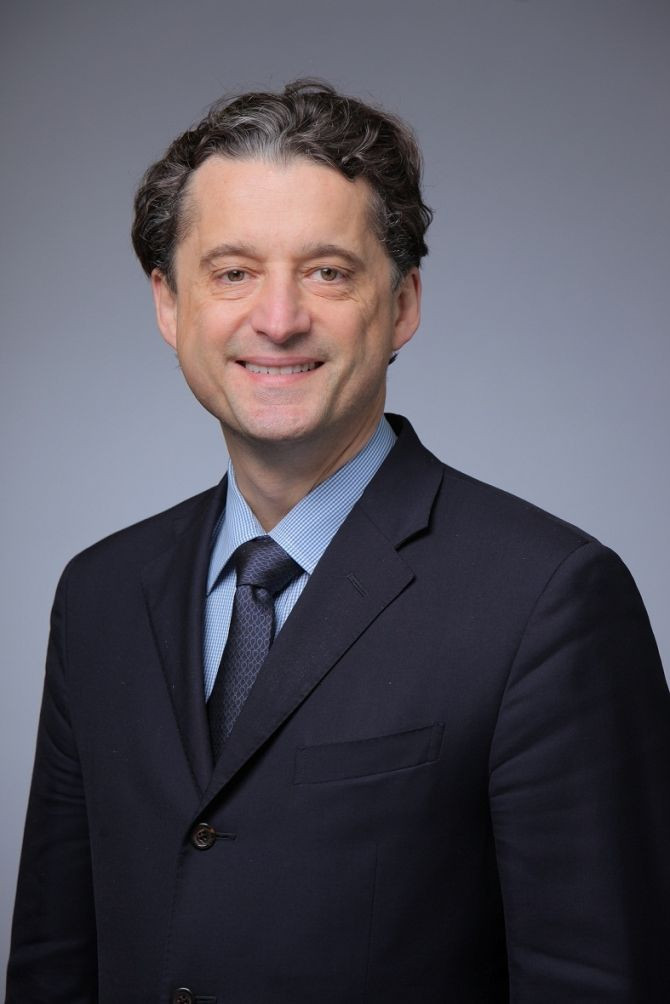
Medical Daily had the chance to talk with the pioneer of robotic-assisted cardiac surgery Dr. Didier Loulmet, Associate Professor and Chief of Cardiac Surgery at NYU Langone Medical Center Department of Cardiothoracic Surgery (CT Surgery).
Dr. Loulmet began to work on minimally invasive cardiac surgery in Paris in 1998.
"We started doing cardiac surgery with da Vinci, we were the first in the world to do mitral valve, endoscopic mitral valve, endoscopic coronary bypass surgery," says Dr. Loulmet. "At the time, the device was a prototype and we saw the value of it. We kept working on it and, today," notes Dr. Loulmet, "It's 14 years later, we are considering this to be one of the best options for patients for cardiac surgery."
Robotic assisted mitral valve repair requires just five small incisions.
"The procedure we most commonly perform is mitral valve repair surgery. Basically, instead of replacing the mitral valve, we repair it," says Dr. Loulmet. "There are techniques for that, that's not new, but the classic operation requires a big incision in the middle of the chest and it's a long recovery from this approach."
According to Dr. Loulmet, "With the da Vinci, we just make five pencil-sized holes on the right side of the chest between the ribs, one hole for the camera, two holes for the instruments, a hole for retraction and a fifth hole for importing and exporting things we need to do the repair."
"But those are very small incisions, five 1cm diameter incisions," notes Dr. Loulmet.
"When the patient heals, six months after surgery, you cannot tell the patient had heart surgery. The advantage is the surgery does not require any retraction on any part of the body to get to the heart, chest wall anatomy is respected, so patients recover much faster with no pain, no nothing."
In 14 years the system is in its fourth generation of development due in part to the many great advances in technology.
"Everything has progressed, the quality of visualization has progressed and the quality of the tools," notes Dr. Loulmet. "Moreover, the da Vinci had three arms, one for the camera and two for the instruments. The fourth arm allows for retraction and exposure and that has been really something that made endoscopic mitral valve surgery possible."
"The precision of the surgery has even gotten better, because of extremely powerful visualization system, with potentially 10x magnification it's like operating with a binocular microscope," says Dr. Loulmet.
The procedure was at first longer than the standard open heart surgery but there was optimism that robotic assisted cardiac surgery was the best option and the advantages of the procedure were readily apparent.
"The procedure lasts around three hours, three and a half hours which is the same as open heart surgery," notes Dr. Loulmet. "In the beginning it used to be a longer operation but now it's the same time. In terms of recovery, the patient spends around three days in the hospital."
NYU Langone has been performing endoscopic mitral valve repair for a year. According to NYU Langone, out of more than 5,000 mitral valve surgeries over the course of 30 years, 4,000 have been mitral valve repairs. Nearly a third of those procedures are now being done robotically.
NYU Langone, where Dr. Loulmet performs the procedure, is the only institution in the tri-state capable of doing so and just one of five in the nation performing the procedure. Expect that to change soon because while there are numerous advantages for the patient, there are just as many for the surgeons.
Robotic assisted surgery goes beyond mitral valve repairs; Dr. Loulmet envisions the future of surgery to be these kinds of robotic assisted procedures.
"I think progressively, in many disciplines, surgery in the future will be performed with this type of robotic-assisted device. It allows for, I think, very strict standardizations of the procedure, it makes it less operator-dependent, says Dr. Loulmet. "It's a different way of operating that requires very tight discipline but when you master the technique, it's really something that you can standardize and can teach in a much easier way than conventional surgery."
As noted by Dr. Loulmet earlier, advancements in visualization technology have allowed for the advances of robotic assisted cardiac surgery and will also shape its future.
Dr. Loulmet's latest research will be aiming to merge what is seen before surgery with what is seen during surgery.
"Because of the progress in pre-operative imaging, we are trying to image the heart with techniques before the surgery so we get a sense of what the lesions are and for that we use a three-dimensional echocardiogram," says Dr. Loulmet.
"And the high quality of visualization, especially when it gets deep inside the heart, we're going to try and correlate the pre-operative imaging and the actual images we get at the time of surgery from the endoscope camera. We are trying to get more experience so we get better and better at pre-operative imaging so we can planify the surgery in a better way. With the correlation between three-dimensional echocardiogram pre-op and what we see inside the heart with the high-magnification visualization system we are trying to optimize the pre-op imaging."
Published by Medicaldaily.com


