Watching Tumors Grow In Real-Time: Researchers Visualize Phases Of Breast Cancer

Researchers from the University of Freiburg in Germany have discovered a new way to watch tumors grow in real-time, testing their method out in rat breast cancer tissue. In their study, they used reflected light to visualize the structure of rat tissue, then used light-based imaging techniques, and fluorophores and dyes, to contrast between the tissues.
Tumor growth is an incredibly complex process, and it’s often difficult to target and treat tumors when they’re constantly changing. As they grow, they alter the microenvironment and cellular composition, the authors explained. Being able to track and monitor these changes on a non-invasive level was the researchers’ aim, as “understanding the temporal changes to the various components within a tumor can aid in the development of improved and customized treatments,” the authors wrote.
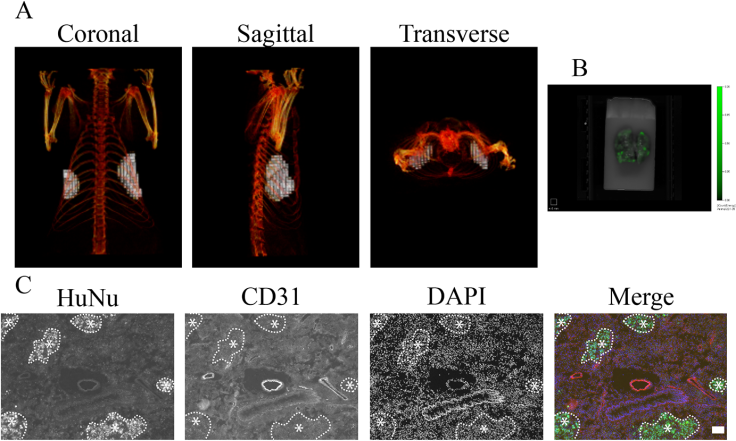
The researchers, led by polymer chemistry professor Dr. Prasad Shastri and pharmacist John Christensen, concentrated on a protein called E2-Crimson, which absorbs light in the far-red spectrum. They were able to track this protein by genetically-engineering it into breast cancer cells, then growing them in a living system. Using infrared light, they illuminated the E2-Crimson in the tumors, and were able to reconstruct a tumor image with fluorescence molecular tomography.
“Non-invasive in vivo imaging is emerging as an important tool for basic and preclinical research,” the authors write in the abstract. “In this study we have developed cell lines stably expressing the far-red fluorescence protein E2-Crimson, thus enabling continuous detection and quantification of tumor growth.”
They found that during the first four weeks that the tumor was developing, the volume increased then later underwent a dramatic increase in the number of tumor cells. “Overall E2-Crimson expressing cells in combination with fluorescence tomography offer a powerful tool for preclinical research,” the authors concluded. “However, further studies in syngeneic tumor models need to be undertaken.”
Source: Christensen J, Vonwil D, Shastri P. Non-Invasive In Vivo Imaging and Quantification of Tumor Growth and Metastasis in Rats Using Cells Expressing Far-Red Fluorescence Protein. PLOS One, 2015.


